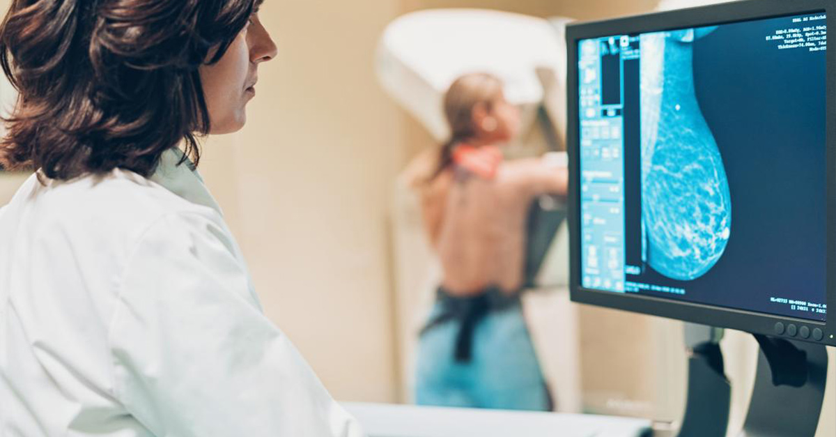
Mammography is a type of medical imaging used to capture images of breast tissue through low-dose X-rays. Doctors may use mammograms to perform routine breast cancer screenings, or to diagnose conditions in women who have been experiencing other concerning symptoms.
The first wave of mammography uses two-dimensional imaging to look at breast tissue. Since its FDA approval in 2011, the 3D mammogram has increasingly become the preferred method in diagnostic centers across the U.S.
What Is a 3D Mammogram?
3D mammography is an advanced form of digital imaging, also known as breast tomosynthesis. The process involves compressing the breast tissue so that the machine can run low-dose X-rays over it. A series of images are then captured by a computer, which assembles the images for a more comprehensive look into glandular breast tissue. As a result, doctors can see breast tissue and any abnormalities in far greater detail than what 2D mammograms can provide.
How Are 3D Mammograms Different from Traditional Mammography?
A 3D mammogram still requires the compression of breast tissue, so the procedure doesn’t differ much from traditional mammograms from the patient’s perspective. Yet, according to experts like Cynthia A. Litwer, MD, chief of breast imaging at Cedars Sinai, and those at Yale Medicine, 3D images are believed to provide better diagnostic results.
Because 2D imaging only captures breast tissue from certain angles — such as the top and side of the breast — tumors and other abnormalities can get hidden in the resulting flat, overlapping images. With 3D mammography, the machine moves along an arc trajectory, capturing 200 to 300 images in total, versus only four captured through traditional mammograms.
With the ability to provide more images in greater detail, 3D mammography can reduce the rate of call-backs for follow-up images. Oftentimes, after a 2D mammogram fails to provide enough detail, patients must go for subsequent imaging to rule out abnormalities. In facilities where 3D mammography is implemented, these patient callbacks can be reduced by 15–30%.
Moreover, the American Cancer Society indicates 3D mammography may be more effective for screening and diagnostics in women with dense breast tissue. In addition to normally occurring milk ducts, glands, and fatty tissue, breasts also comprise supportive, connective tissue. As a result, searching for anomalies can be challenging in areas of density. 3D mammography helps women with dense breast tissue receive even more comprehensive breast cancer screenings.
Better Accuracy = Better Treatment
Because of this more accurate imaging, 3D mammography could detect more cases of breast cancer than 2D imaging.
While research comparing clinical outcomes between the two approaches remains ongoing, even slightly increased odds of detecting breast cancer can be considered a victory. “Currently there is not sufficient knowledge on the causes of breast cancer,” The World Health Organization asserts, “therefore, early detection of the disease remains the cornerstone of breast cancer control.” Oftentimes, mammography can help doctors diagnose cancer before patients can even feel a lump.
Whether you’re due for a breast cancer screening, or your doctor has prescribed a diagnostic mammogram, we encourage you to schedule an appointment with Heartland Imaging for 3D mammography. Our caring team uses the latest technology in mammography to support optimal diagnostic and screening outcomes. Call (502) 429-6500 to schedule your appointment.